
Withering-type botanical microscope, 1780
The “Withering-type Microscope” is named for its inventor, Dr. William Withering (1741-1799), an English physician and botanist who graduated with a degree in medicine 1766 in Edinburgh. Inspired by the taxonomical work and systematic classification of Carl Linnæus (1707-1778), Withering (1776) applied the Linnaean taxonomical system of classification to British plants in a seminal, two volume work, A Botanical arrangement of all the vegetables naturally growing in the British Isles. The earliest reference to a small botanical microscope of Withering’s design appeared in the first edition of this book. There, Withering indicated this microscope was developed for field dissections of flowers and other plant parts. While there is no surviving example of this exact design, close relatives of this type do exist, made either completely of brass or of ivory with brass pillars. Ivory models can be tentatively dated to 1776-1785, as by 1787 a newer model with a hollowed stage in an all-brass configuration already predominated. In turn, it was preceded by the brief appearance of a transitional brass model but with solid stage of ivory or horn (seen here). This version is extremely rare and must have been produced in very small numbers. By 1787 all these varieties were not recorded anymore in the literature.
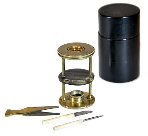
References: SML: A242712; Goren 2014.
References: SML: A242712; Goren 2014.
Prof. Yuval Goren's Collection of the History of the Microscope
Introduction to Polarizing Microscopy
The polarizing microscope is an important tool used in research on materials with crystalline structures. Unlike regular microscopes used for biological studies, a polarizing microscope does more than just make small objects look bigger. It helps to identify what these objects are made of. This microscope is based on a regular light-based microscope, so it can do everything a basic microscope can. However, its ability to examine a material's crystal structure makes it a powerful analytical tool.
When using a polarizing microscope, the definition of matter is based on how light behaves as it moves through a crystal and the colors it creates that we can see. A crystal is a solid piece of matter that has a neat internal arrangement. This neat structure is built from a repeating unit called a unit cell. The way this unit cell is arranged, following specific geometric rules, affects how light travels through the crystal, provided the crystal allows it to pass.
The polarizing microscope has various tools that help focus light and view it in different ways, allowing researchers to determine important properties of the material being examined. These properties help to identify the material and understand what it is made of.

Light behaves in different ways. When light comes from the sun or a light bulb, it spreads out in many directions. This type of light is called "unpolarized light." You can imagine it like waves moving everywhere, without any specific direction. On the other hand, light can also be "polarized." This means all the light waves travel in the same direction, rather than spreading out. You can think of polarized light like waves that move up and down in a straight line, between two points.
To make light polarized, we use a special tool called a "polarizer." When light passes through a polarizer, it only lets the waves that are aligned with it go through. The waves that do not match are blocked. For example, Polaroid sunglasses use this idea to reduce glare from surfaces like water or roads.
The first polarizer was invented by the Scottish scientist William Nicol in 1828. He used two pieces of clear crystal and put them together to filter light. This invention is called a "Nicol prism."
Today, many polarizers are made using something called Polaroid filters. These filters use a special material between two pieces of glass that lets light through only in one direction. This material has tiny crystals arranged in a specific way, creating the Polaroid effect.
When light passes through a polarizing filter, only the waves that align with the filter's polarization can pass; the others are either absorbed or reflected. This makes the light "plane-polarized," meaning that all the waves are aligned in the same direction.
You can think of a polarizing filter like a screen with narrow slits that only lets waves through in one direction. If you have two polarizers turned in opposite directions (90 degrees apart), no light can pass through, and it will be completely dark.
Brewster's Law was discovered in 1811 by a Scottish scientist named Sir David Brewster. This law explains what happens to regular light when it enters a material, like glass. When light goes into this kind of material, it splits into two rays. One ray is reflected (bounces back), and the other is refracted (bends). Both rays become partially polarized, which means they have a specific direction.
Brewster showed that these two rays are most strongly polarized when the angle at which the light hits the surface is the same as the angle at which it is refracted. Usually, when light hits a clear material at an angle other than straight on (90 degrees), it changes direction according to a rule called Snell's Law.
However, when light enters a special kind of material with internal structure, it experiences a phenomenon called double refraction, or birefringence. This happens because of the different forces inside the crystal structure. When light hits the crystal at an angle, it splits into two rays, each polarized in a different direction. These two rays travel at different speeds because they experience different forces from the atoms in the crystal. As they move through the material, they separate from each other. This effect is particularly important for tools such as polarizing microscopes, which help scientists observe details in their samples.
Interference occurs when two or more waves overlap to create a new pattern. This can also happen with light. When two beams of light of the same color meet, they combine to form a single new light ray. This new ray shows how the two wave patterns join together. How these light waves mix depends on where they start, their colors (which we call wavelengths), and how far apart they are. To better understand interference, think about waves on a pond when you throw stones into it. When you throw a stone, it creates waves that move out in circles. If you throw two stones at the same time, they make two sets of waves. These waves move through each other without causing any problems.
When waves from two stones meet, they create a new pattern where they cross. The new pattern's shape depends on the strength of each wave. To find out how strong the combined waves are, you add the strength of each wave together. This idea is called the principle of superposition. It means that the effect on a point in the water from two or more waves is the same as if you added their effects together.
For example, if a point is exactly in the middle of two wave sources, that point will stay still. This happens because the waves arrive at that midpoint simultaneously, canceling each other out. The point where the waves meet is called a point of intersection; at this point, the waves do not cause motion, so there is no wave action.
When two light waves with the same type of movement meet, two things can happen. One is called destructive interference, where the waves cancel each other out. This occurs when waves with the same wavelength meet. The other possibility is constructive interference, in which two different light waves combine to create a new wave. The wavelength of this new wave is the total of the two original wavelengths.
However, you can't see this when light from two lamps meets in a normal room. This is because the lamps do not give a steady flow of light but instead pulse. Since there is always a small time gap between the pulses from the two lamps, they do not mix. For interference to occur, both light rays must come from the same source at the same time, a condition known as coherence.
When two coherent light rays interfere, they can produce both constructive and destructive interference. Destructive interference happens when the waves from both rays arrive at the same time. Constructive interference happens when there is a small delay, and the waves overlap imperfectly.
According to Brewster's law, these effects happen when light passes through certain materials. When light hits a material, some of it bounces back (reflected), and some goes inside (refracted). In thin layers, the light that goes in interacts with the reflected light. This interaction depends on the angle of incidence, the layer thickness, and the materials involved.
If the light source is not monochromatic, meaning it has many different wavelengths, like sunlight or light from a lamp, you will see various colors in different spots. These colors result from interference between light waves of different wavelengths. These are called interference colors, and they depend on how thick the layer is, the materials involved, and the angle at which you see the light.
Early Polarizing Apparatus
The Emergence of Light-Polarizing Apparatus
The essential components of the polarizing microscope—such as Nicol prisms, achromatic lenses, and thin sections—were known as early as the beginning of the nineteenth century. However, it was not until the end of that century that the polarizing microscope became a standard tool in optical mineralogy (AKA petrography). The initial developments occurred in Italy and Britain, but the field reached its peak primarily in France and Germany. This advancement resulted from collaborative efforts among specialists from the Collège de France, the École des Mines, and the Muséum national d'Histoire naturelle, along with skilled instrument builders and the support of Giovanni Battista Amici, the court astronomer of Florence, and later in Germany.
The essential components needed for polarization, particularly the polarization prism named after him, had already been developed by Edinburgh mineralogists William Nicol (1770-1851) and Sir David Brewster (1781-1861) during the early 19th century. In 1834, the Englishman William Henry Fox Talbot (1800-1877) proposed combining Nicol's prisms with a microscope to observe thin sections of rock. In fact, Nicol had suggested this concept a few years earlier. In 1834, Talbot noted remarkable color phenomena created between the crossed Nicol prisms; however, since he was primarily focused on advancing photography—where he is regarded as a key pioneer alongside Louis Daguerre—he did not pursue further research on rocks.
Henry Clifton Sorby (1826-1908) is recognized as the pioneer of optical mineralogy, also known as petrography. An English amateur microscopist and geologist, his major contribution was the development of techniques for thin sectioning of rocks and minerals. By employing polarized light under a microscope, he began producing thin slices of hard rocks, using methods he learned from Professor William Crawford Williamson in zoology. Sorby utilized polarized light to examine these thin, transparent sections of rocks.
In 1858, he published an important memoir titled "On the Microscopical Structure of Crystals." His presentation to the Geological Society of London that same year is now considered the true beginning of microscopical petrography. This presentation ignited a vigorous discussion that remains notable in the society's history. Leonard Horner, the society's president at the time and a friend of Sorby, later recalled that he had never attended a session that generated such skepticism. During this discussion, Sorby was confronted with a quotation from Horace Bénédict de Saussure (1740 – 1799), who had passed away fifty years earlier. Saussure had deemed it ridiculous to study mountains with a microscope. Subsequently, Sorby explored the physical geography of past geological periods, including the wave structures in certain stratified rocks and the origins of slaty cleavage.
From a broad historical perspective, this event can be seen as the defining moment when Britain lost its leadership in geological and mineralogical research—a field it had pioneered in the late 18th and early 19th centuries, thanks in large part to the groundbreaking work of (mostly Scottish) researchers like James Hutton (1726–1797), often regarded as the first modern geologist, and Sir Charles Lyell (1797–1875). Lyell’s 1830 book, Principles of Geology, significantly influenced his student Charles Darwin in formulating the doctrine of uniformitarianism. Other notable Scottish scientists also contributed to this field. William Nicol invented the Nicol prism and developed techniques for making thin sections of minerals, allowing their internal structures to be viewed by transmitted rather than reflected light. Additionally, Sir David Brewster studied the laws of polarization and double refraction in regularly crystallized bodies.

Nörrenberg Polariscope, ca. 1840
Inv. YG-25-005
These developments laid the theoretical groundwork for research in the mid-19th century when Henry Clifton Sorby presented his lecture to the Royal Microscopical Society (RMS). During this time, however, the RMS began to transition into a club for wealthy gentlemen who focused on questions that were largely unrelated to scientific research. There was a growing obsession with expensive and oversized microscopes, with discussions largely detouched from purely scientific questions exemplified by debates over how many lines could be distinguished in Norbert's Test Plate, overshadowing meaningful scientific inquiries.
This trend reflected a broader tendency in Britain to treat research as a form of sport, resulting in the loss of focus on genuine scientific advancement—particularly in the development of the petrographic microscope. This instrument marked an advanced stage in microscope design, functioning as a spectroscope for visible light. In France and the German states, microscopes were traditionally associated with scientific research, rather than being mere status symbols, leading to a less dominant influence of gentlemen's clubs like the RMS.
The "Continental Microscope," as it was referred to by the predominantly Anglocentric historiography of the microscope, was a practical instrument focused on functionality. These economically accessible devices were accompanied by significant investments in optical performance, rooted in scientific research in optics.
© Microscope History all rights reserved

In France, Auguste Bravais (1811-1863) succeeded Victor Le Chevalier as the Chair of Physics at the École Polytechnique in Paris in 1845. He is best known for his research on Bravais lattices, especially his discovery of the 14 unique lattices found in three-dimensional crystalline systems published in a memoir in 1847, which set the theoretical basis for crystallography as a scientific discipline. His successor, later to become the chair of mineralogy at the École des Mines, was Henri Hureau de Sénarmont (1808-1862). It appears that de Sénarmont did not extensively use the microscope in his work. As a skilled mining engineer, his primary focus was on metallic ores. Like many mineralogists of his time, he aimed to determine the optical characteristics of well-formed crystals, known as automorphic crystals, which he isolated from their petrographic context. However, since the microscope is effective mainly for studying thin sections, it did not meet his requirements. Instead, he preferred using a Nörremberg polarization system, created by German physicist Johann Gottlieb Christian Nörrenberg (1787-1862). This system included a polarization source with an adjustable mirror and a short microscope equipped with a Nicol prism, all mounted above a large open plate that could accommodate objects of various sizes. Although the Nörremberg system offers lower magnification than a microscope, it allows for the three-dimensional study of much larger objects—up to several centimeters in size.

Biot's polariscope by Pixii and son, ca. 1838
Nicolas Constant Pixii (1776-1861), and his son, Antoine-Hippolyte Pixii (1808-1835) were at 2, rue du Jardinet, Paris (1818-1838), and later (1838 - 1855) at 18, rue de Grenelle, Saint Germain, Paris.
Inv. YG-23-008
Biot's polariscope is an optical instrument designed to demonstrate and investigate polarization phenomena, particularly the polarization of light through reflection. Developed by Jean-Baptiste Biot, the instrument was created to study Malus's law, which explains how the intensity of polarized light changes as it passes through an analyzer. A typical biot's polariscope consists of a light source, a polarizer (usually made of a stack of glass plates), an analyzer (another polarizing element), and a mechanism for observing the transmitted or reflected light.
It took more than fifty years for the polarizing microscope to become a standard tool in petrography after the Franco-Prussian War of 1870-71. This significant advancement occurred through progress in three interconnected areas:
-
The development of efficient polarizing microscopes that are accessible to petrographers, rather than being limited to the few physicists capable of creating them.
-
The widespread adoption of thin-section preparation, which made it applicable in various laboratories and research centers.
-
An improved understanding and interpretation of the complex optical phenomena that occur when polarized light interacts with crystal structures.

Picart Polariscopes
After the Franco-Prussian War, Émile Bertrand set up his office at 32 Rue Gay-Lussac. Although he never held an official position, he was actively involved in various pursuits, including working as a mineral merchant, writing scientific articles, and translating foreign works. His commercial activities did not diminish his reputation as a respected scientist. Bertrand was a founding member of the Society of Mineralogy, where he frequently published articles and often served as vice president or president. In addition to enhancing polarizing microscopes, one of the notable aspects of Bertrand's extensive work was his design of a large research microscope, which became the preferred instrument among executives at the École des Mines.
After developing his microscopes, Bertrand continued to improve them, particularly focusing on observations made with converging light. He created a specific device that was widely utilized by Ernest Mallard in his theoretical research. The importance of these observations, which help determine the positions of optical axes in crystals, led Bertrand to add an extra lens into the microscope body. This lens is known as the "Bertrand lens" in France and the "Bertrand-Amici lens" in other countries.
The microscope is specifically designed for studying the optical activity of samples, making it highly valuable in the field of mineralogy. It features three essential components: a graduated rotating stage, a polarizer that converts natural light into polarized light (located before the light passes through the sample), and an analyzer that displays the phenomena occurring when polarized light interacts with an object (located after the sample). By rotating the graduated stage, the analyzer's position can be adjusted to determine how much the sample has deviated from the polarization axis, which aids in its identification and characterization.
The main challenge was designing a rotating stage for the microscope that could be accurately turned around its axis, with each objective centered on this axis. To tackle this issue, we have added several individual devices to our collection to investigate the potential for creating an efficient stage that fulfills this requirement..
De Sénarmont passed away prematurely in 1862 and used his Nörremberg microscope only briefly. He was more focused on experimental synthesis than on crystalline optics, which was a research area introduced at the École des Mines by Gabriel-Auguste Daubrée. His successor, François Ernest Mallard (1833-1894), had Émile Bertrand (1844-1909) among his students.


Early Petrographic Microscopes
The first true petrographic microscope by R. Fuess after H. Rosenbusch, Berlin, ca. 1876
It is important to note that Sorby's microscope was not a truly petrographic type. The leading German petrographers of the late nineteenth century, Ferdinand Zirkel (1838-1912) and Harry Rosenbusch (1836–1914), who are recognized as the great masters of systematic petrography, accepted the use of the microscope with reluctance. While it indeed produced striking images, Zirkel noted that it "did not make it possible to identify the main minerals of rocks, such as quartz, feldspar, pyroxene, or amphibole." This limitation is evident in the thousands of pages of their treatises: Zirkel's Lehrbuch der Petrographie (1866) and Rosenbusch's Mikroskopische Physiographie (with the first edition for minerals published in 1873 and for rocks in 1877). Notably, both works contain no illustrations or drawings depicting the textures or arrangements of minerals in rocks as observed under the microscope.
This is the first true microscope for petrographic purposes. Rudolf Fuess, Berlin, made it according to the specifications of Prof. Harry Rosenbusch of Strasbourg.


In the February 1876 publication of the "New Yearbook for Mineralogy," Rosenbusch outlined several requirements for a revolutionary instrument he discussed in his article titled "A New Microscope for Mineralogical and Petrographic Investigations." His demands were as follows:
1) The microscope should allow the examined object to be rotated comfortably in its own horizontal plane while being fixed with crossed Nicol prisms.
2) It must be possible to accurately read the angle of rotation of the object in the horizontal plane.
3) The planes of vibration of the Nicol prisms should have a known position that can be easily restored at any time after any displacement.
4) If sharp adjustment to the maximum extinction cannot be achieved under ordinary white light conditions, there should be convenient options for utilizing sharper methods.
This particular microscope does not have a serial number. According to Prof. Ing. Timo Mappes, it wasn't until the late 1870s that the company's microscopes were consistently signed and numbered. It was purchased from a dealer in California, but its provenance is unclear. It is quite rare to find such a microscope in the USA, as these early petrographic microscopes were primarily German-made and were quickly replaced by newer models by the 1880s after their introduction around 1875. Unless it was brought to the United States by a collector, the only geologist known to have conducted petrographic studies in the USA at that time was Ferdinand Zirkel. He was a professor of geology from Leipzig who was hired by the US government in 1874 to examine the extensive mineral collections gathered during the Geological Exploration of the Fortieth Parallel. However, based on the evidence available, we cannot definitively conclude that this microscope belonged to him.
Petrographic microscope by Constant Vérick, ca. 1885, used by Ferdinand André Fouqué and Auguste Michel-Lévy, pioneers of petrography
This is an unrecorded type of polarizing microscope attributed to Constant Vérick. It was likely made by special order. The microscope features provenance on the stand:
Laboratoire d'histoire
naturelle des corps inorganiques
Collège de France
Or in its full name: Laboratoire de Géologie physique et chimique attaché au cours d'Histoire naturelle des corps inorganiques du Collège de France.
The text originates from the laboratory of Ferdinand André Fouqué (1828 – 1904), who collaborated with Auguste Michel-Lévy (1844 – 1911) on the artificial production of feldspar, nepheline, and other minerals, as well as on meteorites. They produced significant works such as "Minéralogie micrographique: roches éruptives françaises" (1879) and "Synthèse des minéraux et des roches" (1882).
At the age of twenty-one, Fouqué entered the École Normale Supérieure in Paris. From 1853 to 1858, he served as the keeper of scientific collections. In 1877, he became a professor of natural history in the geology chair at the Collège de France in Paris. By 1881, Fouqué was elected a member of the Academy of Sciences and was the first to introduce modern petrographic methods in France.
Fouqué is recognized for his groundbreaking archaeological excavations on the island of Santorini. In 1862, during geological excavations, he uncovered an entire civilization buried beneath a layer of pumice stone, which is believed to have been the result of an eruption around 2000 B.C. His discoveries included walls covered with stucco, decorated with stripes and floral designs, as well as both handmade and wheel-made pottery. These findings are associated with what we now refer to as the Mycenaean civilization ("The Dawn of Greece," The Quarterly Review, 194: 218–243, July 1901; quotes on pp. 219–220). Today, Santorini, also known as Thera, is linked to the Akrotiri culture, which was part of the Minoan civilization of the second millenium B.C.E.


© Microscope History all rights reserved

A microscope comparator for mineralogy and petrology, manufactured by Nachet of Paris, was utilized in this study. The comparator was used in conjunction with the Michel-Lévy interference color chart to identify mineral compositions. This chart was first introduced in 1888 in the book Les Minéraux des Roches, published in Paris. Auguste Michel-Lévy authored the first part of the book, which outlines the methods employed by mineralogists and chemists to examine minerals and utilize the Michel-Lévy Comparator. In Part 2, Michel-Lévy, along with Alf. Lacroix, presents a tabulated account of the physical and optical characteristics of rock-forming minerals. Michel-Lévy acknowledges the contributions of his predecessors, such as F.A. Fouqué, with whom he previously collaborated on Minéralogie Micrographique; Roches éruptives Françaises (1879), which includes an atlas of 55 chromo-lithographed plates. He also acknowledges other notable works, including Des Cloizeaux's Manuel de Mineralogie (1862), Mallard's Traité de Cristallographie (1884), DeLapparent's Cours de Minéralogie (1884), and Rosenbusch's second edition of Mikroskopische Physiographie (1885). Additionally, he references the work of Klement and Renard in Reactions Microchimiques (1886) and their predecessors in microchemistry. However, the most significant contribution is Michel-Lévy's "Tableau Des Biréfringences," which is presented in color for the first time in the 1888 publication.

© Microscope History all rights reserved
J. Swift & Son, Dick Model Petrographic Microscope, 1891, owned by Prof. O.T. Jones, FRS, FGS
For a collector of petrographic microscopes, the Victorian-era microscopes in England are beautiful, but of lesser interest. Although they were provided with polarization equipment early on - for example, since 1870 - and are often offered as polarization microscopes, they are not really. For the salon microscopists, who wanted to see colorful polarization-optical phenomena in the search for the evening kick, they were well suited, but not for quantitative scientific work. An exception is undoubtedly the petrographic microscope by AB Dick, whose principle he described in 1889 and which was conducted in 1891 in the catalogs of the company Swift & Son. It came up with a completely different and innovative technique.
A good review of the Dick microscope and its history is presented by microscope.antiques. For mineralogical work with the polarizing microscope, a defined and precisely measurable rotation of the specimen relative to the position of the polarizers is absolutely necessary. Classically, it uses a graduated turntable, but this means that you have to adjust the mechanical axis of rotation of the microscope stage and the axis of the imaging optics to each other for each individual lens. What today is standard with centering turrets was a major mechanical challenge at the time of the early development of polariscopes, which was solved differently by different companies. AB Dicks innovative approach was a synchronous rotation of the polarizers in a stationary preparation. This was an exactly manufactured, well-stored turntable dispensable and the annoying centering of the lenses especially at high magnification accounted for. Even with the use of additional equipment, such as the universal turntable, this synchronous rotation was advantageous. It was therefore adopted by virtually all the major microscope makers for their large research stands, but mostly in combination with a turntable, so that, depending on the objective, both methods could be used.
The synchronous rotation of the polarizers in the classical Dick microscope is done via a gear transmission and a long pinion rod as coupling between polarizer and analyzer, as it was taken over and improved by Fuess for the large tripod VI. Other companies have developed the principle in the form of closed gearboxes and a better coupling such as Voigt & Hochgesang (later continued by Dr. Steeg & Reuter) and Leitz.
Allen B. Dick first described his design for a polarizing microscope in the Journal of the Royal Microscopical Society (RMS, 1889, pp.432). In this design the stage remains fixed, while the polarizer and analyzer rotate in synchronism by gearing with an angle scale readout on the stage. The microscope was manufactured and signed by J. Swift & Son of 81 Tottenham Court Rd., London (Swift Catalog, 1891). The instrument became known as the Dick Model featuring an English foot with a Traviss rolling slide holder.
This c.1891, English, signed to foot ‘J Swift & Son, London’. There is also the Initials O.T. Jones on top of the box and also engraved on one of the foot stands. Also the word “Lacey” in italics. Standing on large cast brass foot finished in lacquered and anodized brass, trunnions at top support body, large plano-concave mirror on gimbal below substage, substage assembly with rotating Nicol prism on fold out arm, iris diaphragm and focusing condenser all on rotating divided circle for angular measurement, square stage with Swift 2″ patent mechanical stage and main body to rear of stage incorporating the ‘Dick’ rotating mechanism with fine focus via screw and course focusing via diagonal rack work, body tube incorporating a sliding plate with wheel of apertures and slide in/out Bertrand lens, to top a rotating and folding analyzer engraved with 45 degree positions, complete with five Swift objectives, three eyepieces and one blank, bullseye condenser, and other accessories.
Owen Thomas Jones, FRS FGS (1878-1967) was a Welsh geologist. He was born in Beulah, near Newcastle Emlyn, Cardiganshire, the only son of David Jones and Margaret Thomas. He attended the local village school in Trewen before going to Pencader Grammar School in 1893. In 1896 he went up to University College, Aberystwyth, to study physics, graduating in 1900. He then went to Trinity College, Cambridge, and was awarded a B.A. degree in Natural Sciences (geology) in 1902. In 1903 he joined the British Geological Survey, working near his home in Carmarthenshire and Pembrokeshire. In 1910 he was appointed the first professor of geology in Aberystwyth. In 1913 he became professor of geology at the University of Manchester, and then, in 1930, Woodwardian Professor of Geology at the University of Cambridge (until 1943). He dedicated his working life to the study of Welsh geology.
In 1926 he was elected a Fellow of the Royal Society. In 1956 he was awarded the Royal Medal of the Royal Society, and on receiving it he was described as 'the most versatile of living British geologists'. The same year he was awarded the Wollaston Medal and the Lyell Medal of the Geological Society of London. He was twice president of the Geological Society.
He died at the age of 89 having produced more than 140 publications. A year before his death he published a paper describing the Welsh source of the bluestones of Stonehenge (written in Welsh).


.jpg)

Owen Thomas Jones, FRS FGS (1878-1967)

References: MHS 55102,