
Withering-type botanical microscope, 1780
The “Withering-type Microscope” is named for its inventor, Dr. William Withering (1741-1799), an English physician and botanist who graduated with a degree in medicine 1766 in Edinburgh. Inspired by the taxonomical work and systematic classification of Carl Linnæus (1707-1778), Withering (1776) applied the Linnaean taxonomical system of classification to British plants in a seminal, two volume work, A Botanical arrangement of all the vegetables naturally growing in the British Isles. The earliest reference to a small botanical microscope of Withering’s design appeared in the first edition of this book. There, Withering indicated this microscope was developed for field dissections of flowers and other plant parts. While there is no surviving example of this exact design, close relatives of this type do exist, made either completely of brass or of ivory with brass pillars. Ivory models can be tentatively dated to 1776-1785, as by 1787 a newer model with a hollowed stage in an all-brass configuration already predominated. In turn, it was preceded by the brief appearance of a transitional brass model but with solid stage of ivory or horn (seen here). This version is extremely rare and must have been produced in very small numbers. By 1787 all these varieties were not recorded anymore in the literature.
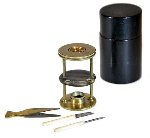
References: SML: A242712; Goren 2014.
References: SML: A242712; Goren 2014.
Prof. Yuval Goren's Collection of the History of the Microscope
Chapter 45: The Microscope in the Crime Scene
The Introduction of Forensic Science
Forensic examination has become an essential component of modern police investigations. Television shows such as CSI and Silent Witness popularized this field in the mid-2000s, significantly broadening its appeal. However, the origins of scientific criminology date back well before the 21st century. Without a real-life Sherlock Holmes in early 1900s France, many of the resources we rely on today may not have been developed.
Dr. Edmond Locard (1877-1966) was a renowned forensic scientist, often referred to as the “Sherlock Holmes of France.” He was born in Saint-Chamond, France, and studied medicine in Lyon. His interests eventually expanded to include the intersection of science and medicine in legal matters.

Edmond Locard and his son in their forensic laboratory
Locard began his professional career by assisting Alexandre Lacassagne, a criminologist and professor. He later partnered with anthropologist Alphonse Bertillon, known for developing a system for identifying criminals based on body measurements. During World War I, Locard served as a medical examiner for the French Secret Service, where he identified the causes and locations of soldiers’ deaths by analyzing their uniforms.
In 1910, the Lyon Police Department provided Locard with the opportunity to establish the first crime investigation laboratory. This lab, located in an unused attic space, allowed him to analyze evidence from crime scenes. Throughout his life, Locard authored numerous publications, with his most famous work being the seven-volume series titled "Traité de Criminalistique" (Treaty of Criminalistics). Locard is recognized as a pioneer in forensic science and criminology. He developed several forensic analysis methods that are still in use today. Notably, he made significant contributions to dactylography, the study of fingerprints. Locard believed that if twelve points of comparison could be identified between two fingerprints, that would be sufficient for a positive identification. This approach became the preferred method of identification, replacing Bertillon's anthropometry.
Locard’s most notable contribution to forensic science is known as “Locard’s Exchange Principle.” According to Locard, “It is impossible for a criminal to act, especially considering the intensity of a crime, without leaving some traces of their presence at the crime scene and taking some traces from this location.” This means that when someone commits a crime, they leave behind evidence of their presence while also taking something away from the scene. Modern forensic science refers to this phenomenon as trace evidence.
The Leitz "COMP" Comparison Microscope for Forensic Science, S.N. 252681, 1927


To accurately compare two objects using optical methods, it is essential to view them simultaneously. This is especially important for small objects that can only be clearly seen under a microscope. When using a single microscope and examining the objects one after the other, it's easy to forget details, leading to potential mistakes, particularly if the objects are similar in color, shape, or size.
This challenge prompted Alexander Alexandrovitch Inostranzeff (1843–1919), a geology and paleontology professor in St. Petersburg, to develop a specialized tool for comparing objects under a microscope. To start his research, he used two single-lens microscopes. One of these was equipped with a "camera lucida," a drawing tool commonly used at the time. By adjusting this drawing device, Inostranzeff could view both objects at the same time, either overlapping or side by side.
While the "camera lucida" improved image clarity, it also had drawbacks. The images produced by the two different microscopes varied not only in size but also in brightness. To resolve these issues, in 1885, Inostranzeff enlisted the help of H. Frantzen, a mechanic at St. Petersburg University, to design a specialized comparison chamber. This chamber could be attached to two nearby microscopes without the need for eyepieces. In this device, light from both microscopes was redirected using mirrors or prisms. The light then traveled to special prisms in the center of the comparison chamber, reflecting the images from each microscope into a single eyepiece tube. At the point where the two prisms converged in the center of the chamber, a dividing line was created, enabling clear comparisons of the images. This comparison chamber served as an effective early version of a device for comparing microscope images.

Alexander Alexandrovitch Inostranzeff (1843–1919) (public domain)

Calvin Hooker Goddard (1891 – 1955) and his comparison microscope (public domain).
In 1911, the Optical Institute of Wilhelm and Heinrich Seibert in Wetzlar developed a groundbreaking type of microscope known as the comparison microscope. By 1917, Ernst Leitz had taken over the design. This innovative microscope featured a unique combination of prisms mounted on a movable platform that could slide from side to side using a screw mechanism. This design allowed users to adjust the alignment between two images, enabling them to view both objects simultaneously with clarity. It was the first microscope to permanently connect two light paths for imaging and included dual illumination, allowing light to be sourced from two angles. Following its creation, Seibert sold the innovative device, and the Wetzlar company received a patent for it.
Around that time, Ernst Leitz also developed the first comparison eyepiece for microscopes. In 1913, he designed the first practical binocular tube that utilized beam-splitting technology, facilitating mass production. As the importance of direct comparison under a microscope became increasingly apparent, by the end of the 1920s, all major microscope manufacturers began incorporating comparison eyepieces into their products.
The challenges of comparing items with a single microscope became evident as laboratories began to focus on accurately identifying forensic bullets. In 1925, the American chemist Philip O. Gravelle conducted the first direct comparisons of bullets using a microscope at the Bureau of Forensic Ballistics in New York. He utilized a special instrument that he had designed himself and later documented his findings.
The Goddard comparison microscope represents a significant achievement in forensic science, celebrated for its innovative design and practical use in criminal investigations. Developed in the mid-1920s by Colonel Calvin Hooker Goddard (1891-1955) and chemist Philip O. Gravelle, this groundbreaking instrument allows investigators to closely examine two specimens side by side—such as a bullet recovered from a crime scene and a test-fired bullet—within a single field of view.
The development of this remarkable device is credited to the partnership between Gravelle and Goddard. Motivated by his doubts about traditional sequential bullet comparisons, Gravelle was the first to conceptualize and create the microscope's initial prototype. Goddard recognized its potential and expertly refined the device, turning it into a reliable forensic tool. A landmark moment in its history occurred during the examination of evidence in the famous Sacco and Vanzetti case in 1927, when the microscope made its debut in forensic analysis. Its reputation grew when Goddard used it in 1929 to analyze the submachine guns linked to the notorious St. Valentine’s Day Massacre, ultimately clearing the Chicago Police Department of suspicion.
These significant investigations led to the establishment of the first independent criminological laboratory at Northwestern University, under Goddard's direction. The design of the comparison microscope is notably intricate, featuring two identical compound microscopes linked by an optical bridge that utilizes mirrors and prisms. This optical connection allows light from both microscopes to merge into a single eyepiece, creating a circular field of view with a central dividing line. To ensure accurate observations, both microscopes must operate under the same magnification and lighting conditions, so any differences noted in the specimens can be attributed solely to the items being examined. Modern versions of this microscope often include advanced translational stages that aid in the precise alignment of matching striae or markings on the bullets being analyzed.
The applications of the Goddard comparison microscope in forensics are extensive and varied. In ballistics, it is an essential tool for comparing distinctive "ballistic fingerprints"—the unique striae left by a gun's barrel—allowing forensic experts to link recovered bullets to specific firearms with impressive accuracy. Beyond ballistics, it plays a critical role in trace evidence analysis, helping forensic scientists identify matches for hair strands, textile fibers, and paint chips found at crime scenes. Additionally, the microscope is invaluable for document examination, helping verify the authenticity of signatures, inks, and typewritten characters. It is also crucial for tool mark analysis, enabling forensic specialists to match tool impressions—such as those made by crowbars or wire cutters—to tools found in a suspect's possession.
Through these various yet vital applications, the Goddard comparison microscope remains an essential tool in forensic investigations, significantly influencing the pursuit of justice and the resolution of criminal cases.


Microscopes in archaeology
Microscopes are essential tools in archaeology, which is the study of past human life. They enable researchers to examine the intricate details of artifacts and materials unearthed from the ground. By using microscopes, archaeologists can gain insights into the composition of these items, their uses, and their origins.
Archaeologists utilize various types of microscopes, ranging from simple light microscopes to more advanced scanning electron microscopes (SEM) that can view extremely small objects. They investigate a wide range of items, including stone tools, pottery, and even ancient plant remains and DNA.
These detailed examinations help archaeologists to better understand history. They can trace how tools were used over time, determine the sources of materials, and gain insights into the diets and environments of ancient peoples.
Key applications of microscopy in archaeology include:
- Use-wear analysis: Studying the microscopic wear patterns on the edges of stone tools can reveal the materials that were cut or processed, such as wood, bone, or meat.
- Material sourcing (provenance studies): By analyzing mineral inclusions in pottery clay or the composition of metal artifacts, archaeologists can identify the origins of these materials.
- Conservation and preservation: High-resolution digital microscopes allow for non-destructive examinations of the surfaces of delicate artifacts, revealing their condition and any signs of corrosion or degradation.
- Analysis of Organic Remains: Microscopic examination can identify small organic residues, such as starch, blood, and hair, that are found on tools and artifacts. This analysis provides valuable insights into how these items were used.
- Identification of Pigments and Materials: Techniques such as Scanning Electron Microscopy with Cathodoluminescence (SEM-CL) effectively identify and map the distribution of various pigments and materials in painted artifacts. This process helps differentiate between similar-looking substances.
For more detailed review of forensic history: