
Withering-type botanical microscope, 1780
The “Withering-type Microscope” is named for its inventor, Dr. William Withering (1741-1799), an English physician and botanist who graduated with a degree in medicine 1766 in Edinburgh. Inspired by the taxonomical work and systematic classification of Carl Linnæus (1707-1778), Withering (1776) applied the Linnaean taxonomical system of classification to British plants in a seminal, two volume work, A Botanical arrangement of all the vegetables naturally growing in the British Isles. The earliest reference to a small botanical microscope of Withering’s design appeared in the first edition of this book. There, Withering indicated this microscope was developed for field dissections of flowers and other plant parts. While there is no surviving example of this exact design, close relatives of this type do exist, made either completely of brass or of ivory with brass pillars. Ivory models can be tentatively dated to 1776-1785, as by 1787 a newer model with a hollowed stage in an all-brass configuration already predominated. In turn, it was preceded by the brief appearance of a transitional brass model but with solid stage of ivory or horn (seen here). This version is extremely rare and must have been produced in very small numbers. By 1787 all these varieties were not recorded anymore in the literature.
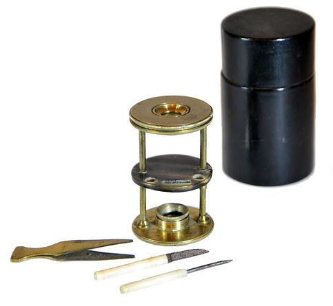
References: SML: A242712; Goren 2014.
References: SML: A242712; Goren 2014.
Prof. Yuval Goren's Collection of the History of the Microscope
Chapter 42: Mid-19th Century Dissecting Microscopes

© Microscope History all rights reserved
Inv. YG-14-003

© Microscope History all rights reserved
Raspail Chemical Microscope, 1835
Initially, based on the aquatic microscope design of the second half of the 18th century, the microscope is mounted on a mahogany box with a drawer and supports a horizontally adjusting eyepiece, a rackwork-adjusted swing-arm stage, and a pivoting mirror; the drawer holds two additional eyepieces and a dissecting set of tools. This microscope was produced by the Parisian optician Louis Joseph Deleuil (1795-1862), according to specifications suggested by François-Vincent Raspail (1794–1878), as a modified version of the Cuff/Ellis type aquatic microscope. In the literature, it is reported to use tourmaline lenses for high magnification (to reduce aberrations, as tourmaline has an extremely high refractive index). Still, my examination of at least the high magnification in this instrument proved it untrue.
This successful design of a portable single microscope was planned by François-Vincent Raspail (1794–1878, picture below with his microscope), a political reformer and activist and a prominent early 19th-century scientist. Raspail (seen in the photo with this type of microscope) was a founder of microscopical methods in chemistry, a pioneer of organic chemistry and one of the first founders of the cell theory in biology. Raspail also pioneered the staining methods in cell biology using iodine to highlight their different parts. As a politician, Raspail was a devoted republican who was active during the restless periods of the First Empire, the Restoration, the Second Republic and the Second Empire, for which he was sentenced several times to prison.
References: SML: 1921-247; Nuttall 1979: 52; Golub: 235; Harvard: 1302, 1905, 1022, 1990-2-0005, 1094; Wissner; Kuhn; Sobel Coll; Balasse; Harvard Univ. 1302, 1095, 1022; Coffeen 2013: 17; Molecular Expressions.

François-Vincent Raspail and his microscope
Hartnack and Oberhauser, Raspail-Type Microscope, 1857-60
This is a later version of Raspail's dissecting microscope, made by George Oberhauser and Edmund Hartnack in Paris.
Edmund Hartnack (1826-91) was a highly esteemed Prussian microscope maker. In 1842-1847 he apprenticed in Berlin under the mechanic Wilhelm Hirschmann senior (1777-1847), which in turn worked with Schiek and Pistor. In 1847 Hartnack moved to Paris to join Heinrich Daniel Rühmkorff (1803-1877), but later he joined the instrument-making firm of Georges Oberhäuser. As opposed to a widespread error probably first introduced by Brian Bracegirdle, Oberhäuser was not Hartnack's uncle! In 1854 Oberhäuser and Hartnack formed a partnership after the latter married Johanna Maria Louise Kleinod, Oberhäuser's niece. In 1864 Hartnack took over the business, from which his former boss had withdrawn more and more. Hartnack established his workshop at 21 Place Dauphin in Paris in 1860. In 1864 he joined forces with the Polish astronomer and mathematician Adam Prazmowski, the former assistant to the Warsaw Observatory and participant in various expeditions to observe solar eclipses and to measure degrees. Prazmowski escaped The January Uprising in Poland and became the production manager of the Hartnack firm. Hartnack had to leave Paris during the French-Prussian War of 1870 and maintained his factory in Potsdam, leaving his partner Prazmowski, in Paris. In 1878, Prazmowski became the owner of the Parisian branch. After his death in 1885, the master Bézu & Hausser took over the workshop and sold it to Alfred Nachet in 1896.


Inv. YG-22-031
© Microscope History all rights reserved
Carl Zeiss, Jena, Dissecting Microscope of the First Model, 1876
Carl Zeiss (1816Carl Zeiss (1816-1888) was born in Weimar as the fifth child of Johann Gottfried Zeiß, a court master turner. After completing his education in Weimar, he apprenticed as a mechanic under Dr. Friedrich Körner, a university mechanic in Jena, from 1834 to 1838. During this time, he also attended lectures at the University of Jena, studying a range of subjects including mathematics, experimental physics, anthropology, mineralogy, and optics. Between 1838 and 1845, Zeiss traveled to various cities, including Stuttgart, Vienna, Berlin, and Darmstadt. In 1845, he returned to Jena and completed an internship at the Physiological Institute under Professor M. J. Schleiden.

On November 19, 1846, Carl Zeiss received a license from the Grand Ducal State Government in Weimar to manufacture and sell mechanical and optical instruments in Jena. He established his first workshop at Neugasse 7 in Jena. In July 1847, the workshop relocated to Wagnergasse 32. That August, August Löber was hired as the first apprentice. Production of simple dissection microscopes began in September 1847. It wasn't until 1857 that the first compound microscopes were produced in the Zeiss workshop. In 1866, the 1,000th microscope was manufactured, marking the beginning of a collaboration with Ernst Abbe. By 1872, the optics of the microscopes had been further refined based on Abbe's calculations.
The earliest Zeiss microscope from 1847/48
The stand is constructed from blackened and varnished brass, black-painted and blued steel, and raw spring steel. For easy packing and portability, the stand can be collapsed into a compact box. It includes a rotating plane mirror to enhance illumination. While earlier models featured a plano-convex condenser lens, this feature has been removed in the current design, as demonstrated in this example.
The microscope comes with all three Chevalier doublets, numbered 1 to 3.
To transport the microscope, it is removed from its walnut case, and the base is placed inside the box in a designated slot to ensure it is stored horizontally. Inside the box, the three original doublets are housed in a small, thick wooden board covered with velvet. A button made of turned white horn (or possibly ivory) allows the board to be lifted, providing access to a compartment underneath for storing the microscope tools.
All parts of the microscope, as well as the wooden case, are marked with a simple “VIII,” indicating that this is likely the eighth instrument in a small series.
In 1849, Zeiss began attaching stands to microscope cases using screws. This design was cost-effective because the mounts could be manufactured on a lathe rather than requiring milling. It also offered practical benefits for users by reducing the risk of the microscope slipping from the guide and falling when placed on the case. A few years after the microscope's introduction, the object clamps on the stand were replaced with versions that were easier to produce.
The early Zeiss dissecting microscope, produced in 1870, features three achromatic doublet lenses that can be used individually or in combination. It also includes an eyepiece lens, allowing for linear magnifications ranging from 8x to 100x. When one or more of the doublets are combined with the eyepiece, they create a Brücke magnifying glass. Additionally, the microscope's table is equipped with leather-covered jaws that support your hands during dissection work.
The "New Dissecting Microscope" was introduced in 1869 as an improvement over the simple dissecting microscopes that had been available since 1847. When this microscope was first described in literature, Carl Zeiss included an illustration of the stand in a woodcut, showcasing its mechanical design, which was similar to the dissection microscope created by Nachet in Paris. This description can be found in Carl Zeiss's 1870 publication, "A New Dissecting Microscope," which appeared in the Archive for Microscopic Anatomy, Volume VI, pages 234-236.
