
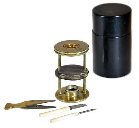
Withering-type botanical microscope, 1780
The “Withering-type Microscope” is named for its inventor, Dr. William Withering (1741-1799), an English physician and botanist who graduated with a degree in medicine 1766 in Edinburgh. Inspired by the taxonomical work and systematic classification of Carl Linnæus (1707-1778), Withering (1776) applied the Linnaean taxonomical system of classification to British plants in a seminal, two volume work, A Botanical arrangement of all the vegetables naturally growing in the British Isles. The earliest reference to a small botanical microscope of Withering’s design appeared in the first edition of this book. There, Withering indicated this microscope was developed for field dissections of flowers and other plant parts. While there is no surviving example of this exact design, close relatives of this type do exist, made either completely of brass or of ivory with brass pillars. Ivory models can be tentatively dated to 1776-1785, as by 1787 a newer model with a hollowed stage in an all-brass configuration already predominated. In turn, it was preceded by the brief appearance of a transitional brass model but with solid stage of ivory or horn (seen here). This version is extremely rare and must have been produced in very small numbers. By 1787 all these varieties were not recorded anymore in the literature.
References: SML: A242712; Goren 2014.
References: SML: A242712; Goren 2014.
Prof. Yuval Goren's Collection of the History of the Microscope
Chapter 39: The Microscope in Uniform
J. Zentmayer, Philadelphia. No. 472
United States Army Hospital Microscope


During the American Civil War (1861-1865), the germ theory of disease was still in its infancy. The link between invisible microorganisms, like bacteria and viruses, and the causes of specific diseases in humans and animals was not well understood. Furthermore, using a microscope to examine the presence of pathogens and detect infections was not a common practice during that time. Still, during the war, microscopes played a role in medical research focused on pathology, wound infections, and blood-borne diseases. Medical microscopes, like the Zentmayer Army Hospital Microscope, were specifically designed for use in military hospitals. The Union Army Surgeon General initially distributed a limited number of microscopes to surgeons, reflecting their scarcity at that time. Although their use was not widespread, these microscopes played a pivotal role in advancing the understanding of infectious diseases in the aftermath of the war. Today, they remain essential tools in modern military medicine.
Joseph Zentmayer (March 27, 1826 — March 28, 1888) was a German American manufacturer of microscopes and optical instruments. After graduating from the gymnasium in Mannheim, he apprenticed with a local optician before working in various optical firms in Germany.
At around age twenty-two, he immigrated to the United States due to his support for the 1848 Revolution. After working in Baltimore and Philadelphia, Zentmayer opened his own shop in Philadelphia in 1858.
During the American Civil War, he supplied microscopes to U.S. government hospitals, beginning production of the U.S. Army Hospital microscope in 1862. This model, known for its size, was mainly used in rear hospital areas for autopsies and microscopic examinations. It featured various innovations patented by Zentmayer, including a swinging substage and a horseshoe foot. By 1879, the basic model was priced at $90, with a fully equipped binocular version at $173.
Zentmayer's craftsmanship is exemplified by his Centennial Model, created for the 1876 exhibition in Fairmount Park, which earned him a medal from the Centennial Commission. He was also recognized with the Elliott Cresson Medal in 1875 and a silver medal at the 1878 Paris Exhibition.
Today, Zentmayer's microscopes are celebrated for their artistry and quality, marking a significant contribution to optical instrument manufacturing in the late 19th century.
George Oberhauser Microscope pour hospices, 1853
Microscopes saw limited use in hospitals by both the Union and Confederate armies during the Civil War, primarily for pathology and the analysis of specimens. At that time, the principles of germ theory were not yet understood. While the Confederacy utilized microscopes to some extent, particularly in larger, rear-echelon hospitals, the Union had a more organized system for procuring and distributing them.
Since the Civil War occurred before the establishment of germ theory, medical professionals on both sides were unaware that microscopic bacteria caused infections and diseases. As a result, practices like sterilizing surgical tools or washing hands between patients were uncommon, contributing to high rates of infection and disease that the microscopes of the era were too weak to study effectively for prevention.
Despite this limited understanding, microscopes were occasionally used for research and diagnostic purposes in select hospitals, rather than in the field, due to their cumbersome nature. They were employed to examine tissue samples, such as bone fragments or tumor-like growths, during autopsies or after surgeries to better understand diseases and injuries. Confederate surgeons like S.E. Habersham used microscopes to study pus from skin lesions in an attempt to investigate complications from spurious vaccinations. Some of these findings were published in medical journals.
The Union Army Medical Department officially published works such as Virchow's Pathology, which addressed microscopic pathology, and commissioned the design and manufacture of a specific "U.S. Army Hospital Microscope" model, making its use more systematic in Union hospitals. Meanwhile, Confederate surgeons had access to both imported European microscopes and domestically manufactured American instruments. Despite facing greater resource shortages due to the Union blockade, the Confederacy managed to utilize these instruments in major hospitals, such as Chimborazo Hospital in Richmond, for specific cases of medical research and diagnostics.
In summary, while microscopes were a novel diagnostic tool during the Civil War, their use was not widespread or consistently applied. The Confederate army mirrored this limited application, focusing on hospital-based analysis rather than battlefield medicine or germ prevention.

References: J. RMS 1900: 379, 381; SML: 1982-250; MHS: 84455; Turner 1989: 200; George; Wissner.

J. Swift & Son, Portable Clinical & Field Microscope, 1900
James Swift and Son designed this collapsing microscope with its solid leather case for the military doctors and traveling scientists at the beginning of the 20th century, undoubtedly under the influence of Charles Baker's "The Diagnostic No. 1" (YG-005 in this collection). At the same time, the same firm designed the more robust "Discovery Microscope" (YG-049) for the Discovery Antarctic Expedition. But the microscope seen here was made to be highly compact and portable without compromising on quality. As the sales advertisement by J. Swift & Son, ca. 1900, states: "This microscope was originally designed to meet the requirements of the Bacteriologist who needed an instrument of utmost portability. It is particularly serviceable to Microscopical and Natural History Societies, as its extreme portability, combined with great steadiness and efficiency for high power investigations, recommends it strongly. These instruments are in very general use in India and Africa amongst those working on Malaria, Sleeping Sickness, etc. They have been supplied in great numbers to the Army Veterinary Departments, the Crown Agents for the Colonies, the United States Government, etc.".


J. Swift & Son, The Discovery Microscope, 1901-10
In 1901 James Swift & Son designed this portable yet competent model specifically for the RRS. Discovery (photo below) for the 1901-1904 Antarctic expedition was the first official British exploration of the Antarctic regions since James Clark Ross's voyage sixty years earlier. Organized on a large scale under the Royal Society and the Royal Geographical Society (RGS) joint committee, the expedition aimed to conduct scientific research and geographical exploration in what was then primarily an untouched continent. It launched the Antarctic careers of many who would become leading figures in the Heroic Age of Antarctic Exploration, including Robert Falcon Scott, who led the expedition, and Ernest Shackleton, who served as the third officer. This microscope was named in the catalog of the time as the 'Discovery' model.
References: J. RMS 1904: 103, 105; MHS: 44459; Billings: P. 111, Fig. 209, AFIP 17783-60-4713-161; Christie's 1995a: 239; Sobel.


