
Withering-type botanical microscope, 1780
The “Withering-type Microscope” is named for its inventor, Dr. William Withering (1741-1799), an English physician and botanist who graduated with a degree in medicine 1766 in Edinburgh. Inspired by the taxonomical work and systematic classification of Carl Linnæus (1707-1778), Withering (1776) applied the Linnaean taxonomical system of classification to British plants in a seminal, two volume work, A Botanical arrangement of all the vegetables naturally growing in the British Isles. The earliest reference to a small botanical microscope of Withering’s design appeared in the first edition of this book. There, Withering indicated this microscope was developed for field dissections of flowers and other plant parts. While there is no surviving example of this exact design, close relatives of this type do exist, made either completely of brass or of ivory with brass pillars. Ivory models can be tentatively dated to 1776-1785, as by 1787 a newer model with a hollowed stage in an all-brass configuration already predominated. In turn, it was preceded by the brief appearance of a transitional brass model but with solid stage of ivory or horn (seen here). This version is extremely rare and must have been produced in very small numbers. By 1787 all these varieties were not recorded anymore in the literature.
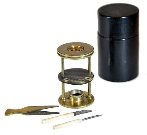
References: SML: A242712; Goren 2014.
References: SML: A242712; Goren 2014.
Prof. Yuval Goren's Collection of the History of the Microscope
Chapter 38: Microscopes by Wilhelm and Heinrich Seibert
Seibert im Wetzlar, Stativ IV (s.n. 5138), 1886
This microscope represents the typical design of the Gundlach-Seibert workshop, featuring a horseshoe-shaped brass base. Rapid movement of the tube through a rack and pinion operated by the knurled knob at the top of the stand (movement without friction); precise adjustment using a fine screw at the bottom operating a parallel linkage system, cylinder aperture with three diaphragms; Concave and plane mirrors movable to both sides.

Inv. YG-22-020
© Microscope History all rights reserved


August Krogh

Ferdinand Julius Kohn

References: Moe 2004, p. 215 Fig. 11.2 (Hans Christian Gram's microscope), pp. 213-220; J. RMS, 1901: 699-701; Boerhaave: V07458; E. Carol. Univ., 48.G29; Wissner.
Model history: Wilhelm (1840 - 1925) and Henry (1842 - 1907) Seibert studied optics under Carl Kellner (1826-1855). After Kellner’s early death, they worked together with Ernst Gundlach and Edmund Hartnack. After acquiring Gundlach's business in Berlin they began the production of high-quality microscopes. The No IV model is based on the original design of Ernst Gundlach, who sold his business to Seibert and Krafft before leaving for the United States. The microscope features the "C limb" and parallel linkage focusing system, typical of several German makers of the end of the 19th century.
The microscope of choice for many researchers: In 1876, Dr. Robert Koch (Nobel Prize in Physiology or Medicine in 1905) purchased a microscope of the IV model seen here for his research of the Anthrax bacteria and photomicrography after advice from his friend and mentor Ferdinand Julius Cohn (1828-1889). During his work, Koch was involved with many microscope improvements in lighting and resolution. He became the first physician to use an oil immersion lens, the Abbe condenser, and photomicrography of bacteria. In his later years, Koch worked with a Zeiss microscope.
Walther Flemming used a Seibert Stativ IV with a second mirror added to illuminate above and below simultaneously for his studies of mitosis. Another microscope of the same model was used by the Danish doctor Hans Christian Gram who developed the staining method called after him. A Seibert microscope of this kind was also employed by August Krogh Bain for his studies of the of the mechanism of regulation of the capillaries in skeletal muscle, for which he was awarded the Nobel Prize in Physiology or Medicine in 1920.

© Microscope History all rights reserved